Human Tooth Structure Vector Diagram.Dental Concept. Education Poster Template, Medical Anatomy
Healthy Tooth Diagram, Tooth Cross Section and Anatomy of Healthy Gum. Medical Dental Poster

Tooth erosion. Tooth erosion is the breakdown and loss of enamel caused by acid or friction. Acidic foods and drinks, can cause it. Stomach acid from gastrointestinal conditions, such as acid.
Tooth Anatomy Carson & Carson, DDS
Parts of a Tooth Diagramed and Explained by a Dentist | Learn the Layers and Tissues of a Tooth FASTParts of a tooth diagramed and explained. This video revi.
Anatomy of the Teeth Dental hygiene school, Dental hygienist school, Dental assistant study

Teeth numbers 1 - 16 are on the upper jaw, also known as the maxillary arch. Teeth number 17 up to teeth number 32 are in the lower jaw, also known as the mandibular arch. Check this printable teeth universal numbering system below. Figure 1. Teeth numbers and names diagram. The human teeth is composed of 16 upper teeth and 16 lower teeth.
Tooth Anatomy Infographic Smile Angels of Beverly Hills
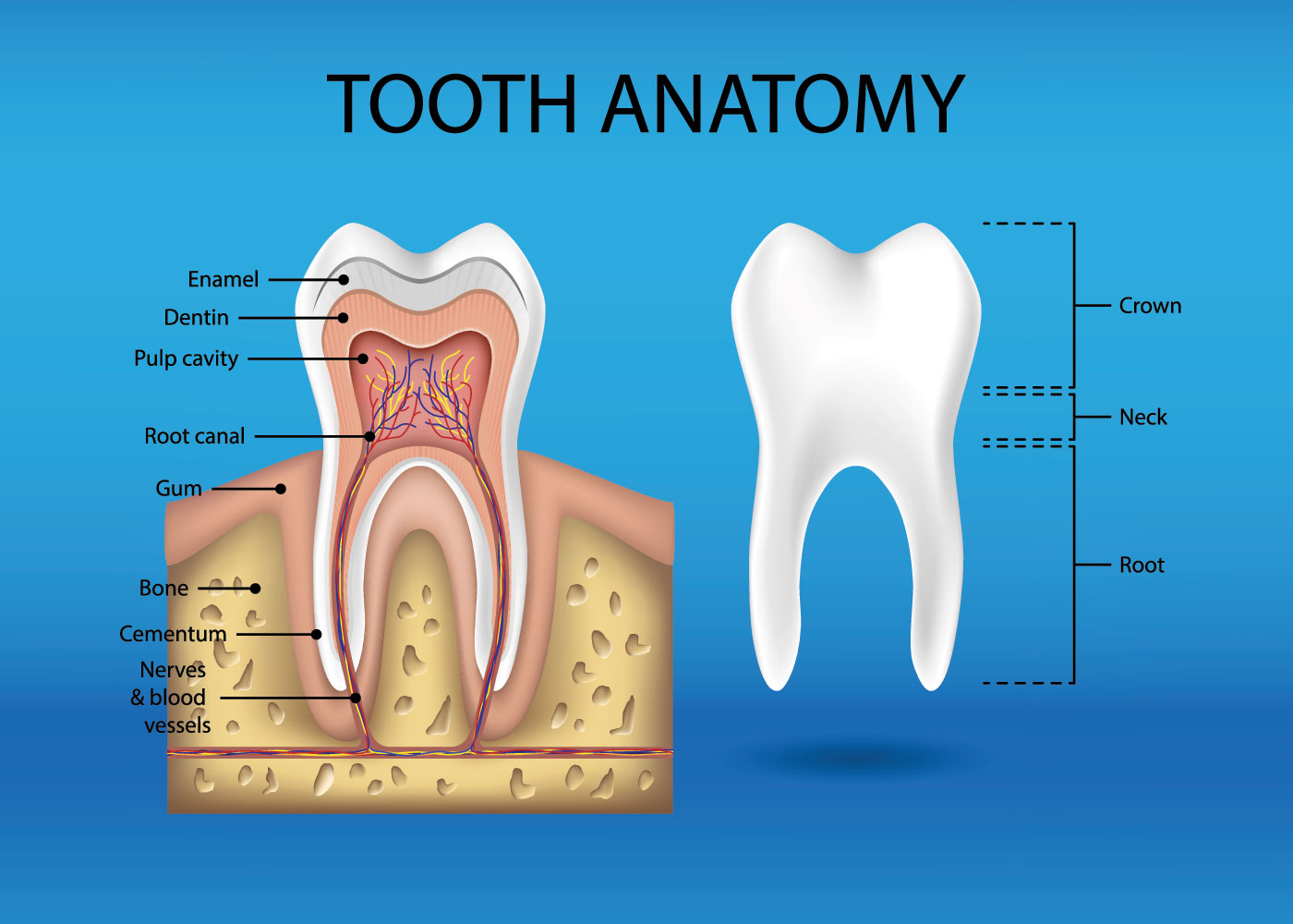
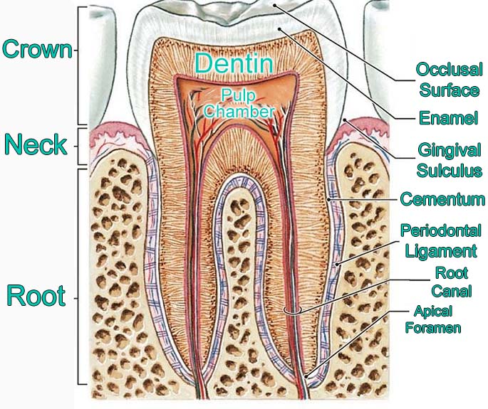
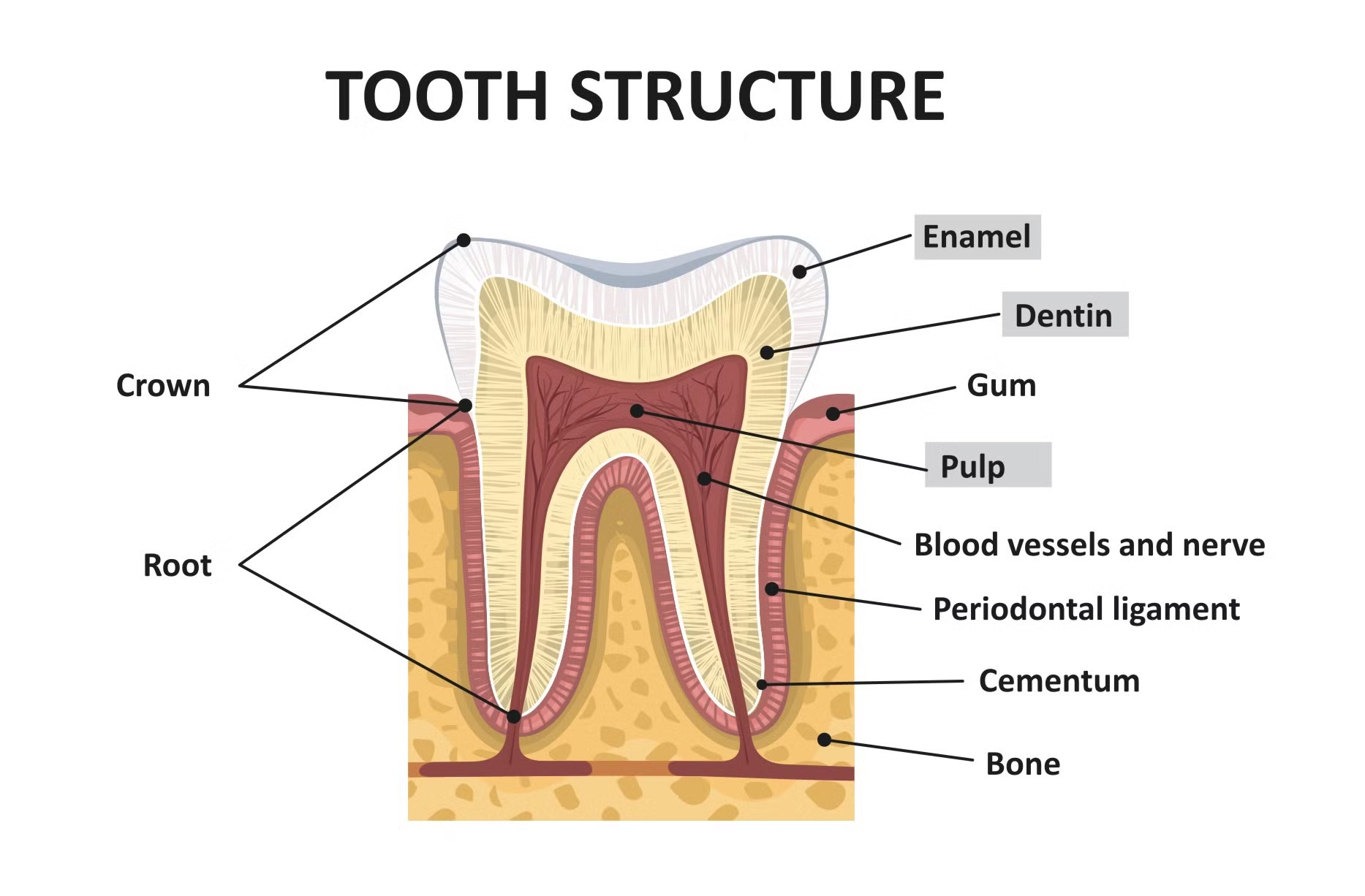
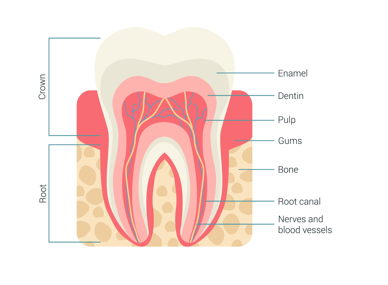
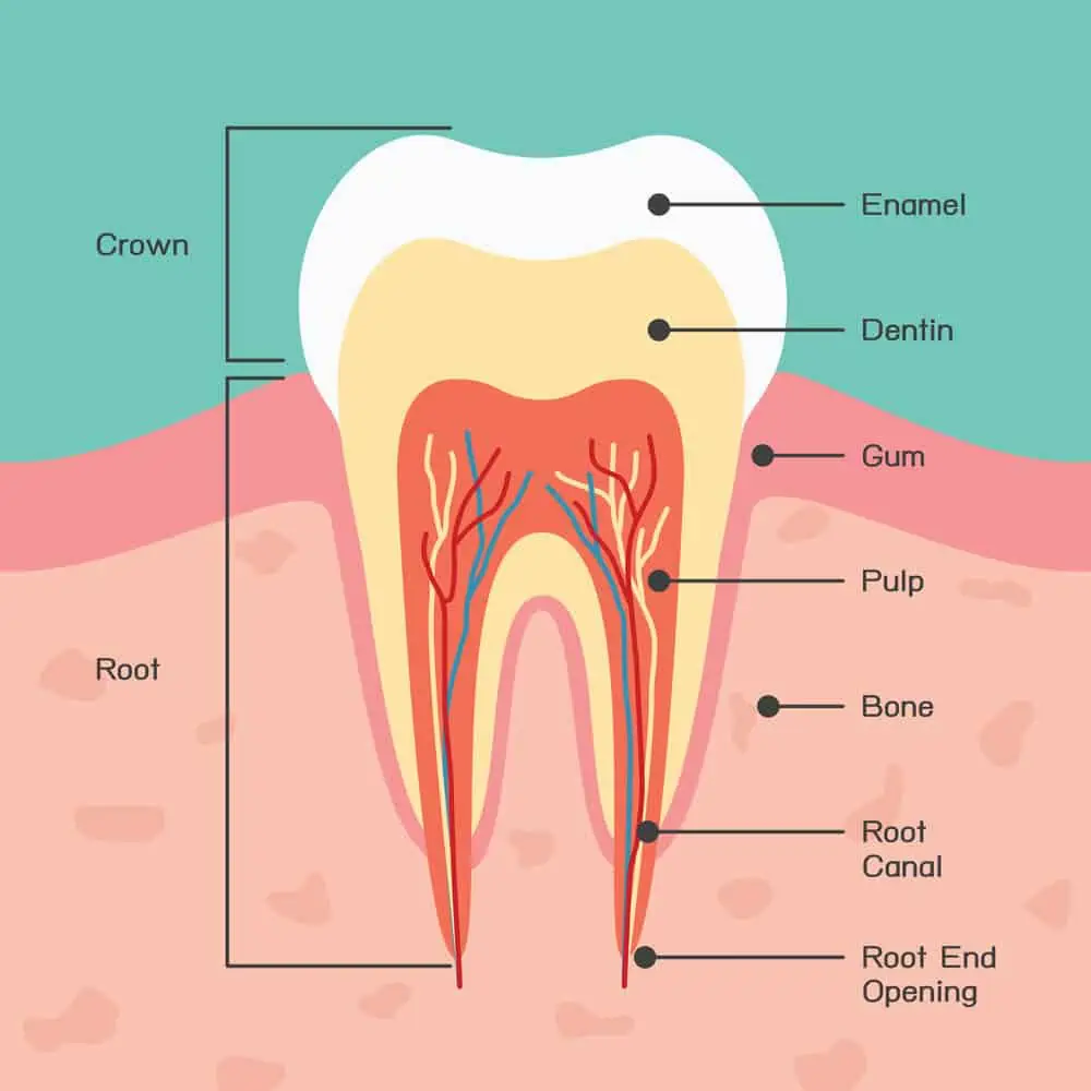
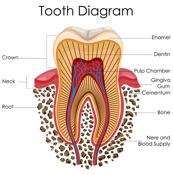
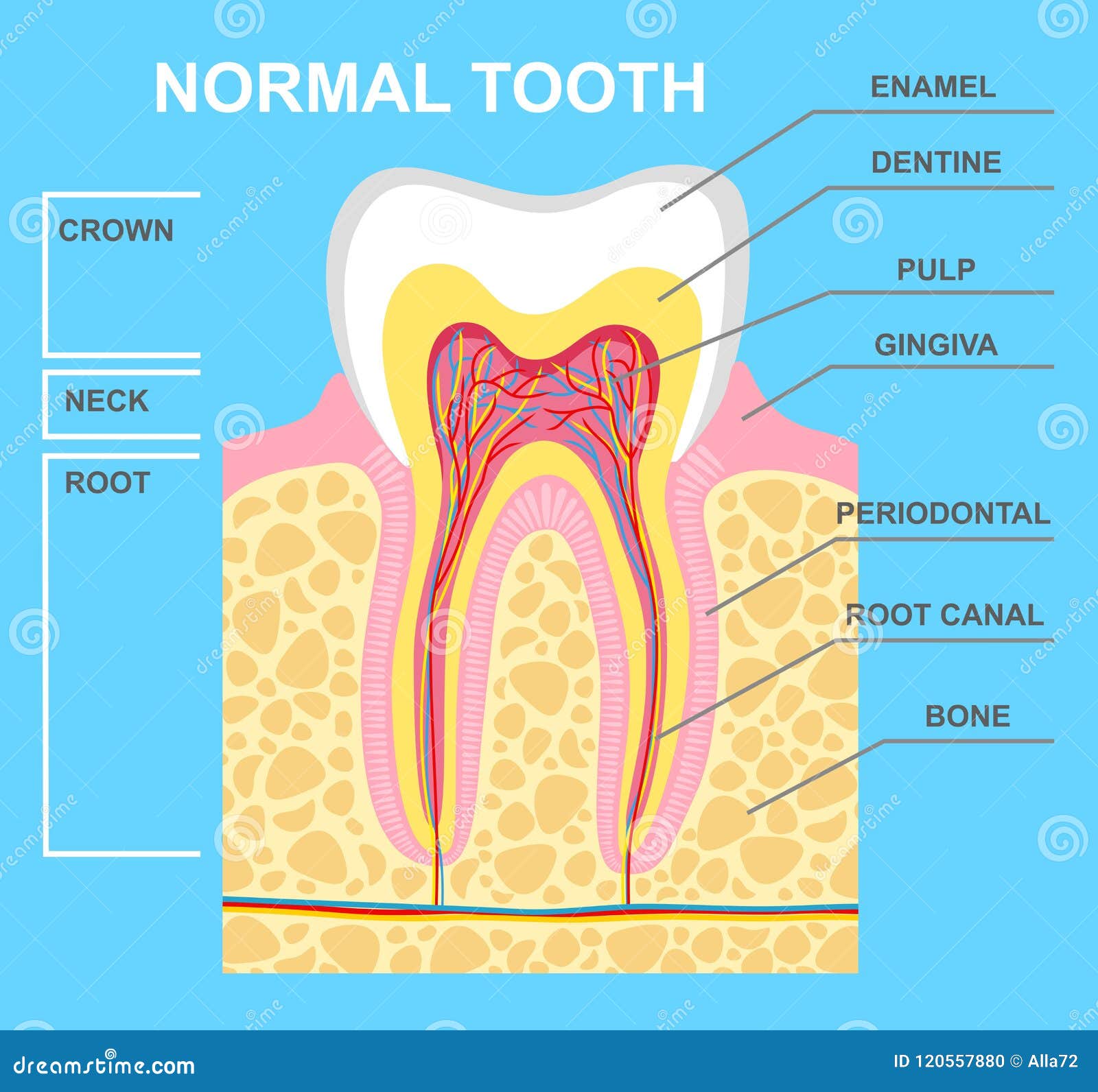
The teeth are a group of hard organs found in the oral cavity. We use teeth to masticate (or chew) food into tiny pieces. They also provide shape to the mouth and face and are important components in producing speech. A tooth can be divided into two main parts: the crown and root. Found above the gum line, the crown is the enlarged region of.
Structure of human teeth Education Illustrations Creative Market
Dental anatomy is a field of anatomy dedicated to the study of human tooth structures. The development, appearance, and classification of teeth fall within its purview. (The function of teeth as they contact one another falls elsewhere, under dental occlusion.)Tooth formation begins before birth, and the teeth's eventual morphology is dictated during this time.
Tooth Anatomy Milford Family Dentistry
Summary. Teeth names include incisors, canines, premolars, and molars. Each type of tooth has a specific function, including biting, chewing, and grinding up food. Teeth are made up of different.
Tooth anatomy
Teeth numbers 1, 16, 17, and 32 are your wisdom teeth (3rd Molars). The teeth numbered 30 and 31 are your lower right molars. If you want to give your smile a new look with dental veneers (Fig. 4), your cosmetic dentist will enhance the most visible teeth in your mouth that always come out when you smile. These are teeth numbers 6 - 11 on the.
Dental tooth anatomy. Cross section of human tooth with infographics and description. 3d
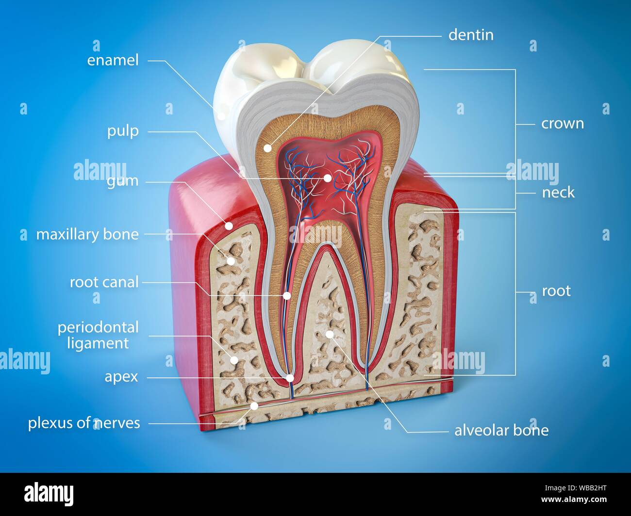
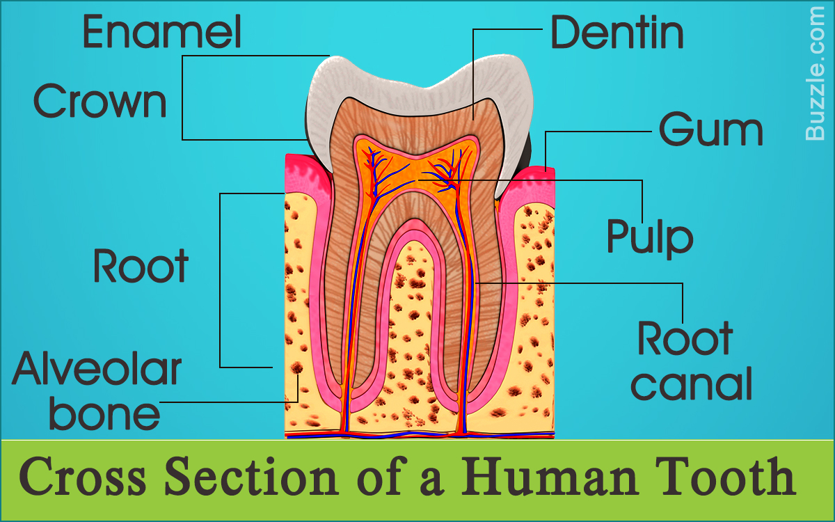
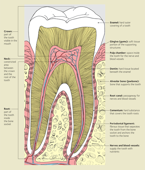
The Anatomy of a Tooth. Your teeth are composed of four dental tissues. Three of them—enamel, dentin and cementum—are hard tissues. The fourth tissue—pulp, or the center of the tooth that contains nerves, blood vessels and connective tissue—is a soft, or non-calcified, tissue. Enamel. Hard calcified tissue covering the dentin in the.
Tooth diagram A Schooner of Science

Human teeth function to mechanically break down items of food by cutting and crushing them in preparation for swallowing and digesting. As such, they are considered part of the human digestive system. Humans have four types of teeth: incisors, canines, premolars, and molars, which each have a specific function.The incisors cut the food, the canines tear the food and the molars and premolars.
Teeth Anatomy Diagram

Your teeth are an essential part of your digestive system. They help you bite, tear and grind food up before swallowing it. To keep your teeth healthy, visit your dentist regularly and practice good oral hygiene at home. With proper care and maintenance, your teeth can serve you well for a lifetime. Medically Reviewed.
Diagram Teeth Roots
There are thirty-two teeth in total in the oral cavity of an adult individual. Sixteen are embedded in the maxilla and sixteen within the mandible.. Look no further than our dental anatomy quizzes and tooth diagrams. Key facts about tooth anatomy; Types of teeth Deciduous dentition (20 teeth), permanent dentition (32 teeth)
Illustration of Human Tooth Diagram. Tooth Structure Vector Illustration Stock Vector
Dental anatomy worksheet activity. An excellent way to revise a new system, organ, region or group of structures is with diagrams labeled with all of the relevant anatomy. By viewing several structures together in one tooth image, you'll be able to: Enter: our labeled tooth diagrams. Spend a few minutes observing the labeled tooth image above.
Set of tooth diagrams 594630 Vector Art at Vecteezy

Tooth, any of the hard, resistant structures occurring on the jaws and in or around the mouth and pharynx areas of vertebrates.. Diagram of human primary and permanent teeth. (more) Like most other mammals, humans have two successive sets of teeth during life. The first set of teeth are called primary, or deciduous, ones, and the second set.
Labeled Teeth Diagram 1 Wiring Diagram Source
Adult teeth are called permanent or secondary teeth: 8 incisors. 4 canines, also called cuspids. 8 premolars, also called bicuspids. 12 molars, including 4 wisdom teeth. Children have just 20.
Digestive System Dental anatomy, Teeth diagram, Human teeth

Crown: This is the top part of the tooth. The shape of the crown enables different functions. For example, the incisors are sharp and are for cutting into food, while the molars have a flat surface for grinding. Gumline: This is where the gum and tooth meet. Root: The root acts as an anchor that keeps your teeth in place.
The Tooth
Types of teeth. The teeth are divided into four quadrants within the mouth, with the division occurring between the upper and lower jaws horizontally and down the midline of the face vertically.. Learn about the types of teeth in a fast and efficient way using our interactive tooth identification quizzes and labeled diagrams.. This leaves up to eight adult teeth in each quadrant and separates.
.