Brain Structure Differentiation Introduction to Neuroscience
Sagittal Section Of Human Brain Labeled Sagittal View Of The Human Brain Labeled Brain diagram

Torcula. Mid-sagittal view of the brain and ventricular system. The medial surface of the left cerebral hemisphere is in view. It is separated from the right hemisphere by the midline falx cerebri (largely removed) occupying the interhemispheric fissure. The lower free edge of this dural fold contains the inferior sagittal sinus, which enters.
Sagittal section of the brain Thalamus, Hypothalamus, Optic chasm, Pituitary gland, Pons

The reticular formation is located throughout the brainstem. Networks within the reticular formation are important for regulating sleep and consciousness, pain, and motor control. The fourth ventricle lies between the brainstem and the cerebellum. Figure 18.2. Regions of the diencephalon and brainstem in a midsagittal section.
Identify the Structures This Midsagittal View of a Brain Model

Download scientific diagram | Mid-sagittal section of the brain with relevant anatomy highlighted. 1 ¼ corpus callosum; 2 ¼ cingulate gyrus; 3 ¼ subcallosal extension of cingulate gyrus; 4 ¼.
MidSagittal Section through the Human Brain by on deviantART Brain

Navigating neuroanatomy can be ridiculously simple if you follow a map. Orient yourself on the main structures on a mid-sagittal section of the brain and the.
Brain Midsagittal View
Lik Thai Lim. Background Cogan's anterior internuclear ophthalmoplegia (INO) is characterized by INO with inability to converge and commonly thought to be due to rostral midbrain lesion. A.
Midsagittal section of the brain anatomy Kenhub
This contains the parts of mid sagittal section of brain as well as the horizontal section of mid sagittal section.
Human Brain Sagittal View

Mid-sagittal Section of the Brain. Anatomy students will be able to identify structures on a dissection. Graphite and Photoshop
LABEL THE BRAIN (MIDSAGITTAL VIEW) Diagram Quizlet

Quick Reference. A cutting through a sagittal plane. The midsagittal section (also called the median section) of the brain passes through the longitudinal fissure and exposes the medial surfaces of the cerebral hemispheres. Compare coronal section, horizontal section, transverse section. From: sagittal section in A Dictionary of Psychology ».
Brain Midsagittal View Diagram Quizlet

BI 335 - Advanced Human Anatomy and Physiology Western Oregon University Figure 4: Mid-sagittal section of brain showing diencephalon (includes corpus callosum, fornix, and anterior commissure) Marieb & Hoehn (Human Anatomy and Physiology, 9th ed.) - Figure 12.10 Exercise 2: Utilize the model of the human brain to locate the following structures / landmarks for the
Sagittal Section Of Brain Diagram Image Of A Longitudinal Section Of A Human Brain The Human

Abstract. The spatial relationships between brain and braincase represent a major topic in surgery and evolutionary neuroanatomy. In paleoneurology, neurocranial landmarks are often used as references for brain areas. In this study, we analyze the variation and covariation of midsagittal brain and skull coordinates in a sample of adult modern.
Brain, Midsagittal View Stock Photo Alamy

⚡ Welcome to Catalyst University! I am Kevin Tokoph, PT, DPT. I hope you enjoy the video! Please leave a like and subscribe! 🙏INSTAGRAM | @thecatalystuniver.
Midsagittal view of brain Diagram Quizlet

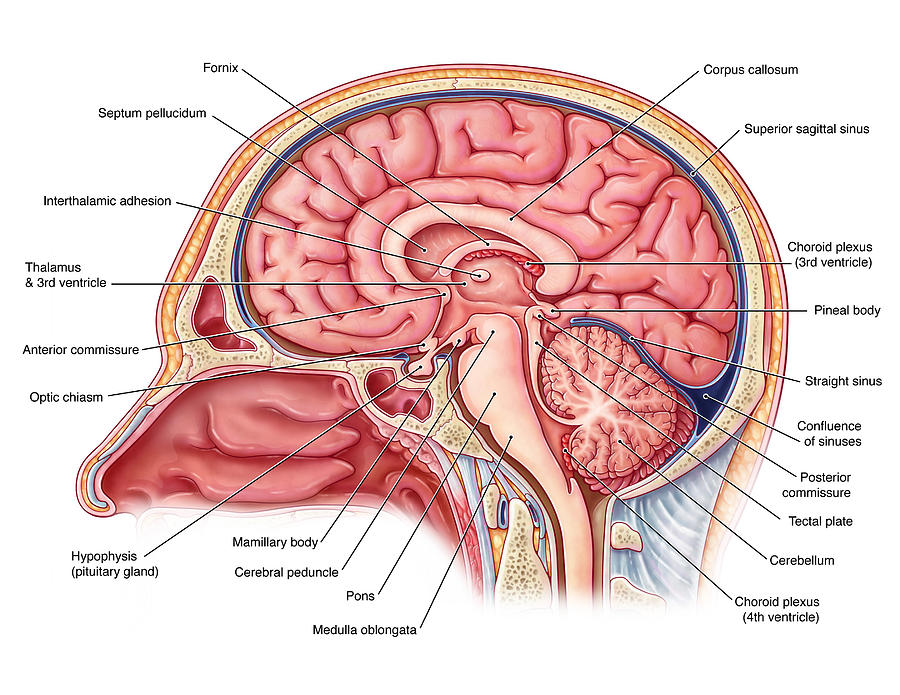
The midsagittal section of the brain shows the three major parts of the brain, which are the cerebrum, cerebellum, and brainstem.The cerebrum (prosencephalon or forebrain) comprises the telencephalon (cerebral hemispheres) and the diencephalon.They are each also divided into subparts or regions for simplified localization of structures, for example, the brainstem is composed of the midbrain.
Sagittal Brain Diagram magicheft
The sagittal midline of the brain is one of the most important sectional planes in neuroimaging. A good working knowledge of the normal neuroanatomy of the sagittal midline is essential so that the subtle abnormalities that may manifest here can be recognized. The neuroembryological development of the brain results in a striking symmetry that.
ANATOMY OF THE MEDIAL (MIDSAGITTAL) SURFACE OF THE BRAIN IN SITU pediagenosis

The midsagittal section of the brain shows the three major parts of the brain, which are the cerebrum, cerebellum, and brainstem. The cerebrum (prosencephalo.
Schematic drawing of a midsagittal view of the human brain. Boxed terms... Download Scientific

Midbrain (Mesencephalon) The midbrain, or mesencephalon, is the most rostral part of the brainstem that connects the pons and cerebellum with the forebrain. For most of its part, the midbrain sits in the posterior cranial fossa, traversing the hiatus of the tentorium cerebelli.. The midbrain is the shortest part of the brainstem. However, it contains many important structures that make it.
Midsagittal Section Of The Brain Color Coded
Structures seen on the medial view of the brain. The images show a midsagittal section of the brain. Watch the video tutorial now.
.